Research Summary: Asymmetry and Dyslexia
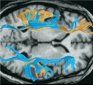
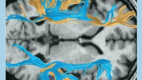
Diffusion Tensor Imaging (DTI) allows scientists to see the white matter fiber tracts that form the major connections among different parts of the brain. Essentially this allows researchers to see a road map of the brain.
There does not appear to be any one pattern for dyslexia, but researchers have found evidence of marked structural differences in dyslexic brains. The image displayed shows a comparison between the brain pathways of a dyslexic man (depicted in blue), and another adult with a very typical brain structure (shown as gold). While the dyslexic subject has less extensive connectivity in the left hemisphere, overall the brain pathways are more evenly distributed and are far more robust in the right hemisphere.
Citation: Asymmetry and Dyslexia, Christiana M. Leonard, Mark A. Eckert, Developmental Neuropsychology, Vol. 33, Iss. 6, 2008
Full Text Article: http://www.ncbi.nlm.nih.gov/pmc/articles/PMC2586924/